SIRT (Selective Internal Radiation Therapy)
Procedure 1: Mapping or Work-up

- The purpose of the angiogram or mapping or work-up is to assess your circulatory system and prepare your liver for the SIRT treatment.
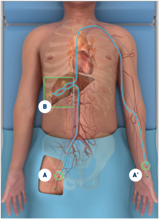
- A small incision is made in a vessel, usually into the femoral artery near the groin (A) or the radial artery (A') near the wrist, and a thin tube (catheter) is threaded through the vessel into the liver (B).
- Your physician will determine which approach is best for your treatment plan.
- The blood vessels are checked on a monitor (angiogram) to make sure that SIRT can be performed safely with the best chance of success.

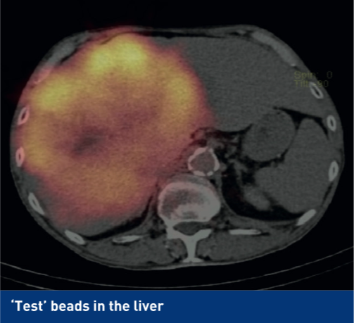
- You will also receive a small amount of radioactive dye or "test beads" to check the potential radiation distribution in the liver, the tumor(s) and other tissue.
- They imitate the radioactive spheres and predict where they will go on the day of treatment.
- The number and location of microspheres following the treatment procedure needs to be predicted. A special scan - named scintigraphy - will be performed to find that out.
Angiogram
The angiogram provides a detailed picture of the blood supply to the liver, which can vary between people. A contrast medium (dye) is injected through the catheter and images of blood vessels are captured using X-rays.
Scintigraphy (lung-shunting scan or MAA scan)
Scintigraphy is an imaging method that uses radioactive materials called radiopharmaceuticals or radiotracers to make things visible. The MAA scan will determine the amount and location of radiotracer absorbed by your body by detecting the radiation from the MAA particles.